
Optomap Retinal Imaging
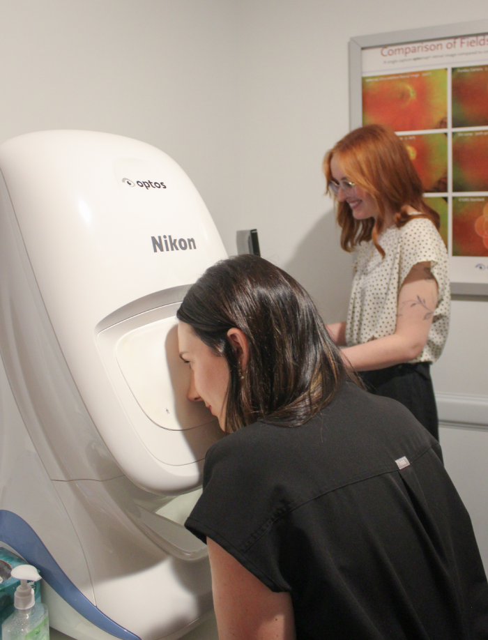
Optomap retinal imaging is a non-invasive technology used in optometry to capture a wide-field image of the retina. This imaging technique provides a detailed view of the back of the eye, including the retina, optic nerve, and blood vessels, without the need for dilation in many cases. It allows optometrists to detect and monitor various eye conditions such as diabetic retinopathy, age-related macular degeneration, and glaucoma. The Optomap system captures a 200-degree digital image of the retina in a single scan, enabling comprehensive examination and documentation of eye health.
Why is retinal imaging our standard of care for all eye exams?
Early Detection Early Diagnosis: This wide-angle imaging can help detect early signs of conditions such as retinal detachment, glaucoma, macular degeneration, and diabetic retinopathy, which may not show symptoms in the early stages.
Non-Invasive and Comfortable Patient Comfort: The procedure is quick, non-invasive, and does not require dilation of the pupils, making it more comfortable and convenient for patients.
Efficient and Effective Time-Saving: The imaging process is fast, usually taking less than a minute, and the images are immediately available for review.
Documentation and Tracking Record Keeping: Optomap images provide a permanent record of your retinal health, which can be compared year after year to track changes and progress.
Education and Communication Patient Education: The images can be shown to patients to help them understand their eye health and the importance of ongoing eye care.

Main
Services
© 2026 Eye Clinic. All Rights Reserved. Accessibility Statement - Privacy Policy - Sitemap